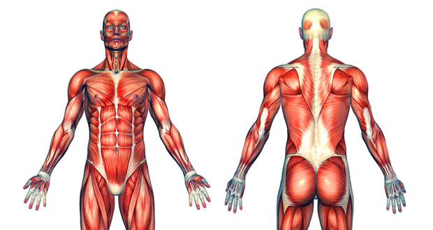
Learn all about human muscles and how they work. Here we explain the major skeletal muscles, muscle structure, fibre types, contractions and sliding filament theory.
Shapes of Skeletal Muscle
What are the different shapes of muscle? There are a number of different muscle shapes within the human body including circular, convergent, parallel, pennate and fusiform. Here we explain where they are found in the body and what their function or purpose is.
Types of Human Muscle
There are three types of muscle found in the human body: Skeletal Muscle Smooth Muscle Cardiac Muscle (heart muscle) Skeletal muscle Skeletal Muscles are those which attach to bones and have the main function of contracting to facilitate movement of our skeletons. They are also sometimes known as striated muscles due to their appearance.
Skeletal Muscle Fibre Types
Within skeletal muscles, there are three types of fibre. Type one (I), type two A (IIa) and type two B (IIb). Each fibre types has different qualities in the way they perform and how quickly they fatigue. Type I Type I fibre are also known as slow-twitch fibre.
Muscle Contraction & Sliding Filament Theory
Sliding filament theory is the method by which muscles are thought to contract. It is recommended that you read the muscle structure page before continuing with the sliding filament theory. The diagram is a common one used to explain sliding filament theory, but don’t worry about trying to understand it all just yet.
Types of Muscle Contraction
Muscle contractions during exercise can be divided into three categories; isotonic (meaning same tension throughout the contraction), isometric (meaning same tension), also known as a static contraction and isokinetic muscle contractions which are performed with a constant speed throughout the movement.
Nerve Propogation & Motor Units
Nerve propagation is the way in which a nerve transmits an electrical impulse. In order to understand this, it is important to understand the structure of a motor neurone (nerve).
Structure Of Skeletal Muscle
Although skeletal muscle cells come in different shapes and sizes, the main structure of a skeletal muscle cell remains the same. If you were to take one whole muscle and cut through it, you would find the muscle is covered in a layer of connective muscle tissue known as the Epimysium.
Major Human Skeletal Muscles
Shoulder Girdle Muscles
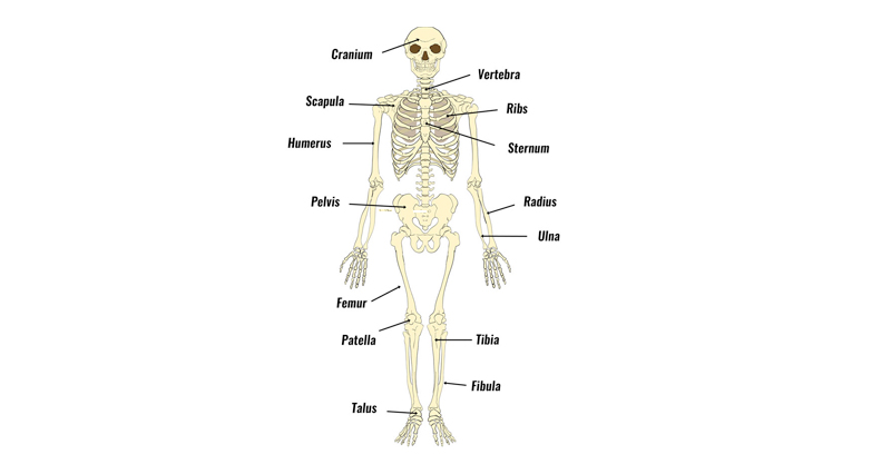
The shoulder girdle consists of the clavicle (collar bone) and the scapula (shoulder blade) which generally move together as a unit. Only the clavicle connects directly to the rest of the skeleton at the sternum bone. It is really only the scapula that moves from action of the muscles.
Shoulder Joint Muscles
The shoulder joint, also known as the glenohumeral joint is a ball and socket joint and consists of the humerus (upper arm bone), clavicle (collar bone) and scapula (shoulder blade). The muscles which stabilize and enable movement of the joint are the pectoralis major, teres major, supraspinatus, deltoid and latissimus dorsi.
Elbow Joint Muscles
The elbow joint consists of the humerus (upper arm bone), radius and ulna in the forearm. The ulna is the bone on the little finger side of the forearm (remember l in ulna for little finger) and the radius radiates around it. The muscles at the elbow joint are the biceps brachii, brachialis, brachioradialis, triceps brachii (triceps muscle), anconeus, pronator teres, pronator quadratus, and supinator.
Wrist and Hand Muscles
The main muscles which move the wrist and hand consist of the flexor carpi radialis, palmaris longus, flexor carpi ulnaris, extensor carpi ulnaris, extensor carpi radialis brevis, extensor carpi radialis longus, flexor digitorum superficiialis, flexor digitorum profundus, flexor pollicis longus, extensor digitorum, extensor indicis, extensor digiti minimi, extensor pollicis longus, extensor pollicis brevis and adductor pollicis muscle.
Thigh & Knee Muscles
The knee joint consists of the femur (thigh bone), tibia and fiblua bones of the lower leg and the patella or kneecap. The muscles which flex and extend (bend and straighten) the joint are the quadriceps muscles (rectus femoris, vastus lateralis, vastus medialis) and the hamstring muscles at the back of the thigh (semitendinosis, semimembranosus, and semitendinosis).
Hip and Groin Muscles
The main muscles of the hip and pelvis consistsof the iliopsoas, pectinues, rectus femoris and sartorius at the front. The gluteus medius, gluteus minimus, piriformis, tensor fasciae latae on the outside. Gluteus maximus, biceps femoris, semitendinosus, semimembranosus at the back and the adductor or groin muscles (adductor brevis, adductor longus, adductor magnus and gracilis).
Lower Leg and Ankle Muscles
The muscles of the lower leg consist of the gastrocnemius and soleus muscles which together are known as the calf muscles, the peroneus longus, peroneus brevis, extensor digitorum longus, extensor hallucis longus, tibialis anterior, tibialis posterior, flexor digitorum longus and flexor hallucis longus.
Neck and Back Muscles
Major muscles of the neck and back include the erector spinae, multifidus, rectus abdominus, transversus abdominus, internal obliques, external obliques, splenius and quadratus lumborum.


