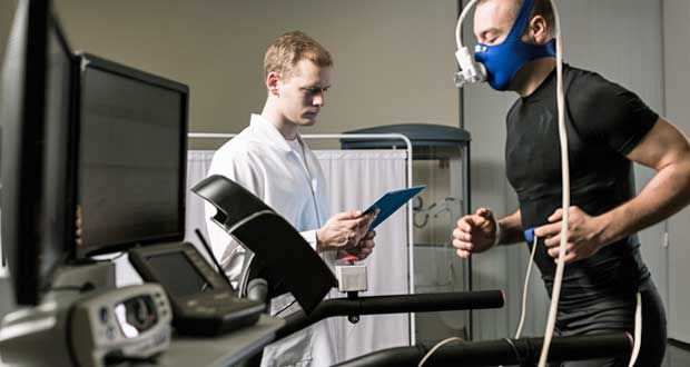
The action of breathing in and out is due to changes in pressure within the chest (thorax). This action is also known as external respiration and is created by the muscles of the chest and the diaphragm changing the size of the chest cavity (and air pressure). Here we explain the mechanics of breathing and how breathing is regulated at rest and during exercise.
Mechanics of breathing
When we inhale the intercostal muscles (between the ribs) and diaphragm contract to expand the chest cavity. The diaphragm flattens and moves downwards and the intercostal muscles move the rib cage upwards and out. This increase in size decreases the internal air pressure and so air from the outside (at a now higher pressure than inside the thorax) rushes into the lungs to equalise the pressures.

When we exhale the diaphragm and intercostal muscles relax and return to their resting positions. This reduces the size of the thoracic cavity, thereby increasing the pressure and forcing air out of the lungs.
Breathing Rate
The rate at which we inhale and exhale is controlled by the respiratory centre, within the Medulla Oblongata in the brain. Inspiration occurs due to increased firing of inspiratory nerves and so the increased recruitment of motor units within the intercostals and diaphragm. Exhalation occurs due to a sudden stop in impulses along the inspiratory nerves.
Our lungs are prevented from excess inspiration due to stretch receptors within the bronchi and bronchioles which send impulses to the Medulla Oblongata when stimulated.
Breathing rate is all controlled by chemoreceptors within the main arteries which monitor the levels of Oxygen and Carbon Dioxide within the blood. If oxygen saturation falls, ventilation accelerates to increase the volume of Oxygen inspired.
If levels of Carbon Dioxide increase a substance known as carbonic acid is released into the blood which causes Hydrogen ions (H+) to be formed. An increased concentration of H+ in the blood stimulates increased ventilation rates. This also occurs when lactic acid is released into the blood following high-intensity exercise.
Regulation of breathing
Respiration is controlled by the autonomic nervous system, which enables us to alter our breathing without thinking about it. The autonomic nervous system consists of two branches, the sympathetic nervous system (the pedals) and the parasympathetic nervous system (the breaks).
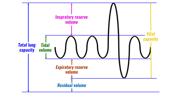
At rest, we inspire approximately 500 ml of air per breath and on average we breathe 12-15 times per minute. The volume of air we breathe in or out per breath is known as tidal volume, and the volume of air we breathe in or out per minute is known as minute ventilation.
Minute ventilation is calculated as follows:
Minute ventilation = tidal volume (500 ml) x breathing rate (15) = 7500ml/min (7.5 l/min)
Our respiration is coordinated by the respiratory centre in the medulla oblongata of the brain. The medulla oblongata could be considered “the boss” and controls many important functions in the body. During inspiration (breathing in), nerve impulses are sent via the phrenic and intercostal nerves which stimulate the inspiratory muscles, the external intercostal and diaphragm, causing them to contract, this stimulation lasts for approximately two seconds, after which, the inspiratory muscles relax and expiration occurs. Expiration is a passive process
Regulation of breathing at rest
- Medulla oblongata controls breathing
- Phrenic and intercostal nerves stimulate the external intercostal muscles and diagram
- Stimulation causes these muscles to contract
- Contraction of these muscles results in inspiration
- Stimulation ceases, muscles relax and expiration occurs
- Regulation of breathing during exercise
During exercise, a significant rise in minute ventilation occurs, this is due to an increased oxygen demand from the working muscles. Both tidal volume and breathing rate increase to compensate for an increased oxygen demand, therefore increasing minute ventilation.
Central to the increase in rate and depth of breathing during exercise are a series of receptors. Of particular importance are the chemoreceptors, which are found in the aortic arch and carotid arteries and detect blood acidity. Chemoreceptors detect blood acidity by monitoring the concentrations of oxygen and carbon dioxide. During exercise, the chemoreceptors detect a rise in carbon dioxide, a by-product of increased respiration, and a reduction in oxygen.
The chemoreceptors, send a nerve impulse to the medulla oblongata, which subsequently stimulates the sympathetic nervous system (the pedals) to increase breathing rate and depth.
Proprioceptors detect movement in the joints and muscles. During exercise, the proprioceptors detect a rise in movement and therefore oxygen demand, and send a nerve impulse to the medulla oblongata, which stimulates the sympathetic nervous system to increase breathing rate and depth.
During exercise, the depth of breathing is increased through the stimulation of three additional muscles. In addition to the external intercostal muscles and diaphragm, the sternocleidomastoid, scalene, and pectoralis minor are stimulated to lift the ribs and sternum further, increase the volume of the thoracic cavity, allowing an increase in the depth of breathing.
Breathing frequency is also increased during exercise due to the expiratory centre being activated and stimulating the expiratory muscles, the abdominals, and internal intercostal muscles, making expiration an active process and increasing the rate of expiration. Stretch receptors prevent over-inflation of the lungs, so if the lungs are excessively stretched, the expiratory centre sends impulses to induce expiration.