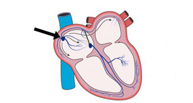
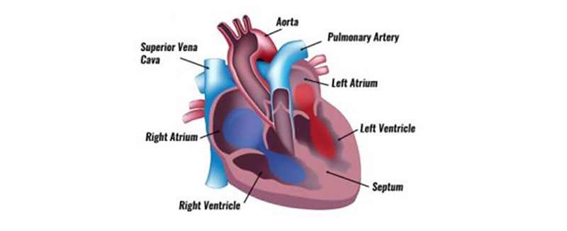
The heartbeat is caused by impulses arising from two specialised groups of cells within the heart muscle. The Sino-Atrial (SA) node, situated in the wall of the right atrium initiates the beat, and the Atrioventricular (AV) node which is positioned between the ventricles and continues to distribute the wave of impulses.
The heartbeat
There are several distinct stages that form a full heartbeat. Cardiac Systole describes the period at which the heart contracts and cardiac diastole describes the period of relaxation, between beats. They can, however, be further divided into diastole and systole of the atria and ventricles.
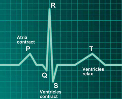
The phases of a heartbeat can also be divided into sections relating to the shape of the electrical signals produced when viewing the heartbeat via an ECG (Electrocardiogram). This traces the electrical activity of the heart. The wave shape produced is called the QRS wave, with each part of the wave being labeled to help describe what is happening at each stage.
- TP Interval (Ventricular Diastole) – Atria and ventricles are relaxed, and blood is flowing into the atria from the veins. As the atrial pressure increases above that of the ventricle, the AV valves open, allowing blood to flow into the ventricle
- P Wave (Atrial Systole) – The SA node fires and the atria contract causing atrial systole which forces all blood into the ventricles, emptying the atria.
- QR Interval (End of Ventricular Diastole) – The AV valves remain open as all remaining blood is squeezed into the ventricles. The electrical impulse from the SA node reaches the AV node which spreads the signal throughout the walls of the ventricles via specialised cells called bundles of His and Purkinje fibres. The R peak is the end of ventricular diastole and the start of systole.
- RS Interval (Ventricular Systole) – As the blood is now all within the ventricles and so the pressure is higher here than in the atria, the AV valves close. The ventricles start to contract although pressure is not yet high enough to open the SL valves
- ST-Segment (Ventricular Systole) – Pressure increases until it equals Aortic pressure when the SL valves open. The blood is ejected into the Aorta (and pulmonary artery) as the ventricles contract. At this time the atria are in diastole and filling with blood returning from the veins.
- T Wave (Ventricular Diastole) – Ventricles relax, the ventricular pressure is once again less than the aortic pressure and so the SL valves close. The cycle continues.

